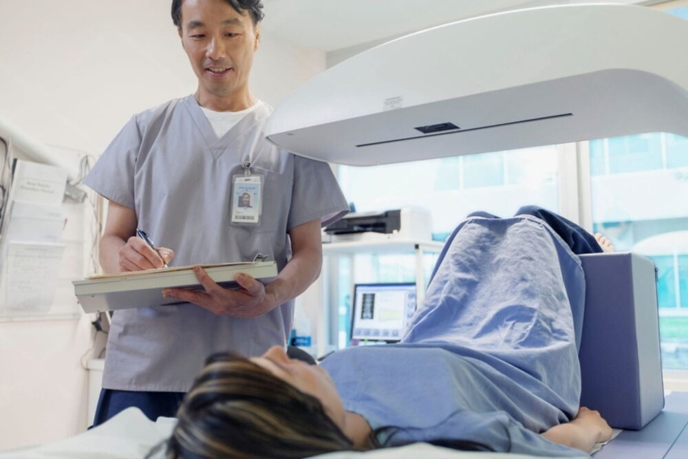
Accurate diagnosis is the foundation of effective orthopaedic care. Bones, joints, muscles, ligaments, and nerves work together in complex ways, and many injuries or conditions cannot be fully understood through symptoms alone. For individuals in Mansfield, Fort Worth, and Burleson, TX, medical imaging plays a critical role in clarifying what is happening beneath the surface. Lone Star Orthopaedic and Spine Specialists, PLLC emphasizes education around diagnostic tools so patients can better understand how specialists evaluate musculoskeletal conditions. Imaging supports orthopaedic diagnosis, and different imaging methods can contribute to a clearer clinical picture.
Why Imaging Is Essential in Orthopaedics
Orthopaedic symptoms such as pain, stiffness, swelling, or weakness can stem from a wide range of causes. Two people with similar symptoms may have very different underlying conditions. Imaging allows specialists to visualize internal structures that cannot be assessed through physical examination alone, helping to distinguish between soft tissue injuries, bone changes, joint degeneration, or nerve involvement.
Without imaging, diagnosis would rely heavily on assumptions based on symptoms and movement patterns. While physical evaluation provides valuable information, imaging adds precision. It helps confirm suspected conditions, rule out others, and guide next steps in evaluation or management. This clarity is especially important when symptoms persist, worsen, or involve multiple structures.
Common Types of Orthopaedic Imaging
Several imaging modalities are commonly used in orthopaedics, each offering unique insights. X-rays are often the first imaging tool used to evaluate bone structure, alignment, fractures, and joint space changes. They are particularly useful for identifying arthritis, fractures, and deformities.
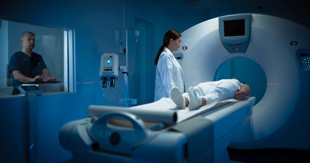
Magnetic resonance imaging (MRI) provides detailed images of soft tissues such as muscles, ligaments, tendons, cartilage, and nerves. MRI is valuable when evaluating complex joint injuries, disc issues in the spine, or unexplained pain where soft tissue involvement is suspected. Computed tomography (CT) scans offer detailed cross-sectional images of bones and are often used for complex fractures or surgical planning.
Ultrasound is another imaging option that allows real-time visualization of soft tissues and joint movement. It can be useful for assessing tendon injuries or guiding certain procedures. Learning about orthopaedic imaging helps individuals understand why different tools are selected based on the suspected condition.
How Imaging Supports Accurate Diagnosis
Imaging helps confirm what specialists observe during physical evaluation. For example, joint pain accompanied by reduced range of motion may suggest arthritis, but imaging can reveal the extent of cartilage loss or bone changes. Similarly, persistent pain after an injury may raise concern for ligament or tendon damage, which imaging can help identify.
In spine-related conditions, imaging plays a particularly important role. Symptoms such as radiating pain, numbness, or weakness may be linked to disc changes or nerve compression that cannot be detected externally. Imaging allows specialists to correlate symptoms with structural findings, supporting a more precise diagnosis.
Imaging and Injury Severity
Not all injuries are immediately apparent. Stress fractures, small ligament tears, or early degenerative changes may not cause dramatic symptoms at first. Imaging can reveal subtle abnormalities before they progress, helping explain why pain persists even when external signs are minimal.
Understanding injury severity also supports appropriate expectations. Imaging findings can clarify whether a condition is mild, moderate, or advanced, which helps individuals understand why symptoms behave a certain way. This knowledge supports informed decision-making and reduces uncertainty about what is happening inside the body.
The Role of Imaging in Chronic Conditions
Chronic orthopaedic conditions often involve gradual structural changes rather than sudden injury. Arthritis, tendon degeneration, and disc wear develop over time and may fluctuate in severity. Imaging helps document these changes and provides context for symptom patterns.
For individuals with long-standing joint pain, imaging can reveal whether symptoms are related to joint space narrowing, bone spurs, or soft tissue inflammation. Educational resources related to general orthopaedic health often highlight how imaging supports understanding of chronic joint conditions and their progression.
Imaging in Spine and Neck Evaluation
The spine is a complex structure that protects the spinal cord while allowing flexibility and movement. Imaging is essential when evaluating neck or back symptoms that involve neurological changes, such as tingling, numbness, or weakness. MRI and CT scans provide detailed views of discs, vertebrae, and nerve pathways.
Understanding spinal imaging helps individuals appreciate why certain symptoms extend beyond the neck or back. For example, arm or leg symptoms may originate from spinal structures rather than the limb itself. Learning about neck and spine anatomy provides helpful context for how imaging supports accurate diagnosis in these cases.
Imaging and Treatment Planning
While imaging itself does not determine treatment, it provides essential information that informs next steps. Understanding the location, extent, and nature of an injury or condition helps specialists consider appropriate options within a broader care plan. Imaging findings are interpreted alongside symptoms, physical examination, and functional limitations.
It is important to note that imaging findings do not always correlate perfectly with pain. Some individuals have imaging changes without significant symptoms, while others experience pain with minimal visible changes. This is why imaging is used as part of a comprehensive evaluation rather than in isolation.
Addressing Common Misconceptions About Imaging
One common misconception is that imaging always provides a definitive answer. While imaging offers valuable insight, it is one piece of the diagnostic process. Another misconception is that more imaging is always better. In reality, the type and timing of imaging are carefully considered to avoid unnecessary exposure or confusion.
Education helps individuals understand why imaging may or may not be recommended at a given time. Clear communication about the purpose of imaging supports realistic expectations and reduces anxiety around diagnostic findings.
How Imaging Improves Patient Understanding
Beyond diagnosis, imaging plays an important role in patient education. Visualizing internal structures helps individuals better understand their condition and why certain symptoms occur. Seeing images of joints, bones, or soft tissues can make abstract explanations more concrete.
This understanding supports engagement in care decisions and encourages adherence to recommended activity modifications or rehabilitation strategies. When individuals understand what is happening internally, they are often better equipped to participate actively in their own musculoskeletal health.
The Bigger Picture of Musculoskeletal Health
Orthopaedic imaging supports understanding not only of isolated injuries, but of how different parts of the body interact. A foot issue may influence knee alignment, while shoulder mechanics may relate to neck posture. Imaging helps reveal these relationships by showing how structures align and function together.
Educational information related to ankle and foot mechanics can further illustrate how lower extremity alignment influences overall movement patterns and joint health.
Learn More About Orthopaedic Imaging
Imaging is a powerful tool that enhances accuracy, clarity, and understanding in orthopaedic diagnosis. By revealing internal structures and supporting precise evaluation, imaging helps specialists better understand musculoskeletal conditions and explain findings clearly.
Lone Star Orthopaedic and Spine Specialists, PLLC provides educational insight into orthopaedic diagnosis and imaging for individuals in Mansfield, Fort Worth, and Burleson. Understanding the role of imaging empowers individuals to approach evaluation with confidence and clarity, supporting informed decisions about musculoskeletal health.
Sources
Modic, M. T., & Ross, J. S. (2007). Lumbar degenerative disk disease. Radiology.
Reinus, W. R., & Khurana, B. (2014). Imaging of musculoskeletal injuries. New England Journal of Medicine.
Hunter, D. J., & Bierma-Zeinstra, S. (2019). Osteoarthritis. The Lancet.