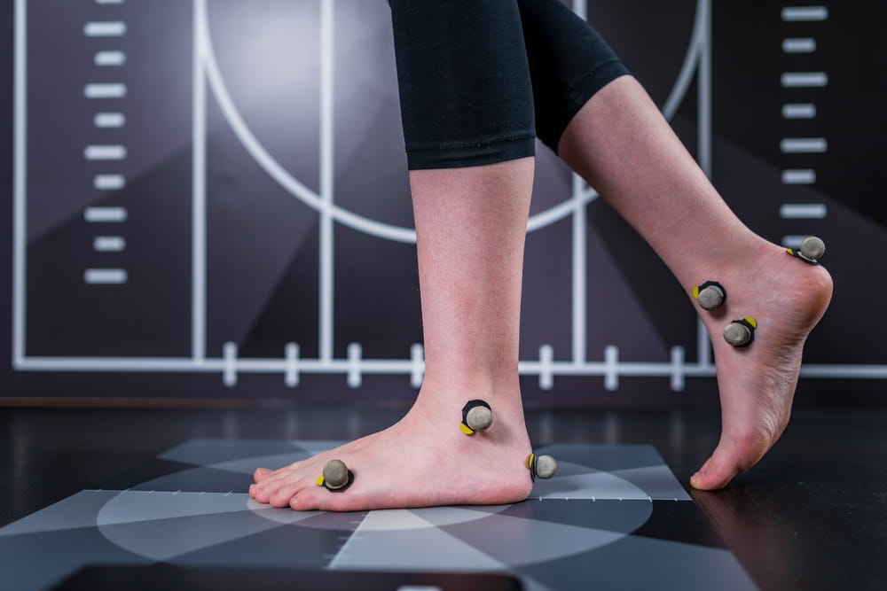
A painful joint can provide plenty of clues without immediately revealing exactly what has been injured. Swelling, tenderness, limited motion, weakness, instability, or difficulty bearing weight may suggest damage to a bone, ligament, tendon, cartilage surface, or another structure. A physical examination helps narrow those possibilities, and imaging may provide additional information when more detail is needed.
Different imaging tests answer different questions. An X-ray is often useful for evaluating bones and joint alignment, while MRI can provide a closer look at many soft tissues. Ultrasound and CT also have specific roles depending on the injury and the part of the body involved.
Lone Star Orthopaedic and Spine Specialists, PLLC uses examination findings and appropriate orthopaedic imaging to evaluate joint injuries for patients throughout Mansfield, Fort Worth, Burleson, and surrounding North Texas communities. The goal is not simply to order the most advanced scan available, but to select imaging when it can help clarify the diagnosis or guide treatment.
Imaging Is One Part of an Orthopaedic Evaluation
A scan is rarely the first piece of information a clinician considers.
The evaluation usually begins with questions about how the injury occurred. A twisting injury may suggest a different group of problems than a direct impact, fall, or sudden pull. The clinician may also ask whether the joint swelled immediately, whether the patient heard a pop, and whether the joint feels unstable or locks during movement.
The physical examination adds more information by assessing tenderness, range of motion, strength, stability, swelling, and the ability to perform certain movements.
Imaging is then used when the history and examination suggest that seeing the structures inside the joint could change the diagnosis or treatment plan.
Why X-Rays Are Often the Starting Point
X-rays are among the most commonly used imaging studies after a joint injury because they provide detailed information about bones.
If someone falls on a wrist, twists an ankle, or lands directly on a knee, an X-ray may help identify a fracture or dislocation. It can also show whether the bones remain properly aligned.
X-rays may reveal arthritis, bone spurs, narrowing of joint spaces, or previous injuries that could also be contributing to symptoms.
However, X-rays do not show every type of injury. Ligaments, tendons, muscles, cartilage, and many other soft tissues are not visualized with the same level of detail.
That limitation does not make an X-ray less useful. It simply means that another type of imaging may be appropriate when the suspected injury involves soft tissue rather than bone.
When MRI Provides More Detail
Magnetic resonance imaging, or MRI, creates detailed images of both bone and many soft tissues without using ionizing radiation.
MRI is particularly useful when an orthopaedic clinician suspects damage to structures such as ligaments, tendons, cartilage, muscles, or the meniscus in the knee. It can also help identify some bone injuries that are not obvious on initial X-rays.
For example, after a knee injury, an MRI may help evaluate a suspected ACL tear, meniscus injury, cartilage damage, or other internal joint problem. In the shoulder, MRI may provide information about the rotator cuff, labrum, or surrounding tissues.
An MRI is not automatically necessary for every painful joint. If the diagnosis is already clear from the history, examination, and X-rays, additional imaging may not change treatment.
CT Scans and Complex Bone Injuries
Computed tomography, commonly called CT, uses X-rays taken from multiple angles to create detailed cross-sectional images.
CT can be especially helpful when a fracture is complex or when the clinician needs a clearer view of how broken pieces of bone are positioned around a joint.
For certain injuries involving the wrist, ankle, hip, or other complicated areas, CT may provide information that is difficult to obtain from standard X-rays alone.
Because CT uses ionizing radiation, it is generally selected when the additional detail is likely to influence treatment or surgical planning rather than used routinely for every joint complaint.
Ultrasound Can Show Structures in Motion
Musculoskeletal ultrasound uses sound waves to create real-time images of soft tissues.
One advantage is that a tendon or other structure can sometimes be observed while the patient moves. This can be useful when evaluating certain tendon injuries, muscle problems, cysts, or fluid around a joint.
Ultrasound may also be used to guide injections so medication can be placed accurately around a specific joint or soft-tissue structure.
Its usefulness depends partly on the location of the injury and the expertise of the person performing the examination. Deep structures and certain internal joint problems are usually better evaluated with other forms of imaging.
Different Joint Injuries Require Different Tests
There is no single imaging study that is best for every injury.
A clinician may consider several factors before choosing a test:
- How the injury occurred
- The location of pain and tenderness
- Whether the patient can bear weight or use the joint
- How much swelling or instability is present
- Whether a fracture is suspected
- Whether symptoms suggest ligament, tendon, cartilage, or muscle damage
- Results from any previous imaging
- Whether the test is likely to change treatment
This is why two patients with pain in the same joint may receive different imaging recommendations.
Knee Injuries
The knee contains bones, ligaments, cartilage, tendons, and menisci, making it a common example of how different imaging methods complement one another.
After significant trauma, X-rays may be used first when the examination suggests a possible fracture or other bone injury. If the bones appear intact but the knee remains unstable, locked, or significantly swollen, MRI may be considered to evaluate internal soft tissues.
A twisting injury during sports, for example, may injure the ACL or meniscus without producing an obvious abnormality on an X-ray.
Physical findings remain important because an MRI can reveal abnormalities that are not necessarily responsible for the patient’s current symptoms.
Shoulder Injuries
The shoulder’s wide range of motion depends heavily on soft tissues for stability.
X-rays may help identify fractures, dislocations, arthritis, or changes involving the bones and joint spaces. MRI or ultrasound may provide more information when a rotator cuff tendon injury or another soft-tissue condition is suspected.
Imaging selection also depends on whether symptoms developed after a traumatic event or gradually through repetitive use.
Someone who suddenly loses the ability to raise the arm after a fall may need a different workup than someone experiencing mild shoulder discomfort that gradually developed over several months.
Wrist and Hand Injuries
Falls onto an outstretched hand frequently lead to wrist injuries.
X-rays are often used first to look for fractures. However, some wrist fractures can be difficult to identify immediately, particularly when they are small or nondisplaced.
If pain and tenderness remain highly suspicious despite normal initial X-rays, repeat imaging or another study may be considered.
MRI, CT, or ultrasound may also be appropriate when a ligament or tendon injury is suspected. Current imaging guidelines emphasize matching the next study to the particular clinical concern rather than automatically ordering the same test for every patient.
Ankle and Foot Injuries
Ankle injuries range from uncomplicated sprains to fractures, cartilage injuries, and significant ligament damage.
The physical examination helps determine whether imaging is needed. When a fracture is suspected, X-rays are typically the initial test.
If persistent pain, instability, or other symptoms continue despite apparently normal bone imaging, additional testing may help evaluate ligaments, tendons, cartilage, or subtle bone injuries.
Because many ankle sprains improve without advanced imaging, MRI is generally reserved for cases in which symptoms, examination findings, or the course of recovery raise additional concerns.
Imaging Can Reveal Findings That Are Not Causing Pain
One of the most important things to understand about orthopaedic imaging is that an abnormal scan does not automatically identify the source of symptoms.
Age-related changes, minor disc abnormalities, cartilage wear, and tendon changes may appear on imaging in people who have little or no pain.
This is why imaging must be interpreted in context. A finding matters most when it matches the patient’s symptoms, mechanism of injury, and examination.
Ordering imaging without a clear clinical question can sometimes uncover unrelated changes that complicate rather than clarify the diagnosis.
Why an MRI Is Not Always the First Step
Patients sometimes assume that an MRI is the most complete test and therefore should be performed immediately.
In reality, the most useful test depends on what the clinician is trying to identify. If the primary concern is a fracture, an X-ray may provide the information needed more quickly and efficiently.
In some injuries, a period of appropriate treatment may also be reasonable before advanced imaging is considered. If symptoms improve as expected, additional testing may not be necessary.
If pain persists, function worsens, or examination findings suggest a more significant soft-tissue injury, MRI may become more useful.
Imaging May Help Guide Treatment
Once a diagnosis is established, imaging can help determine the severity of an injury and whether different treatment options should be considered.
A small, stable injury may be treated differently from a displaced fracture or complete ligament tear. Imaging can also help clinicians determine whether surgery should be considered or whether nonsurgical treatment remains appropriate.
In many cases, treatment includes activity modification, medication, bracing, and rehabilitation. Physical therapy may be used to restore motion, strength, stability, and confidence as the joint heals.
Follow-up imaging is not always necessary. Clinical improvement in pain, strength, motion, and function may provide sufficient evidence that recovery is progressing appropriately.
When Joint Injuries Need Prompt Evaluation
Some injuries should be evaluated promptly rather than monitored at home for an extended period.
Concerning signs include an obvious deformity, inability to bear weight or use the affected limb, severe swelling, loss of normal joint motion, or significant pain after trauma. Numbness, weakness, unusual skin color, or loss of circulation beyond the injured area may require urgent attention.
A joint that repeatedly locks, gives way, or remains significantly swollen after an injury also deserves evaluation.
The appropriate imaging study, if one is needed, can then be selected based on the suspected injury.
Using Imaging to Complete the Clinical Picture
Orthopaedic imaging is most valuable when it answers a specific question. X-rays can provide important information about bones and joint alignment. MRI can reveal many soft-tissue injuries. CT offers detailed views of complex bone anatomy, while ultrasound can evaluate selected structures dynamically.
None of these tests replaces a careful history and physical examination. Instead, imaging adds another layer of information when symptoms or examination findings require greater detail.
Across Mansfield, Fort Worth, Burleson, and the surrounding North Texas region, Lone Star Orthopaedic and Spine Specialists, PLLC uses imaging as part of a broader approach to joint injury evaluation. Connecting the images with the way an injury happened, the patient’s symptoms, and the physical examination can lead to a more focused diagnosis and an appropriate treatment plan.
Sources
American College of Radiology. ACR Appropriateness Criteria: Acute Trauma to the Knee.
American College of Radiology. ACR Appropriateness Criteria: Acute Hand and Wrist Trauma.
American Academy of Orthopaedic Surgeons. Orthopaedic Imaging and Diagnostic Guidance. OrthoInfo.